Electroencephalogram (EEG)
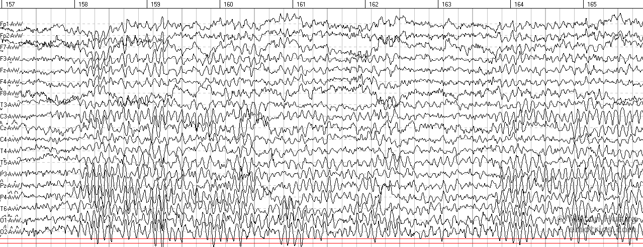
The electroencephalogram or EEG is a measurement of the electrical activity of the brain, obtained by placing multiple electrodes on the scalp. The use of multiple electrodes covering the entire head creates some ability to localize the origin of different electrical signals.
In medical settings, EEG is most commonly used by neurologists to understand seizure activity in the brain or to see if there is a lack of activity and brain death. The EEG is also used in sleep studies to identify the different stages of sleep.
For studying the brain, EEG has advantages of high temporal resolution, able to detect changes on a scale of milliseconds, as compared to fMRI which has a resolution on the scale of seconds. It is also more portable. A MRI machine requires a one ton (or more) magnet (a 3T magnet weighs 30,000 lbs). On the other hand, MRI has the advantage of high spatial resolution, able to detect physical brain distortions in the range of millimeters (tenths of mm for the big magnets). EEG can localize electrical sources with centimeter accuracy depending on the number of electrodes that are used.
Most clinical EEG machines use 19 amplifier channels to measure signals from 19 electrodes arranged according to the standardized 10-20 system, named for the relative spacing across the head (10%-20%-20%-20%-20%-10%) between electrodes. This standardization allows for 'caps' to be made with all the electrodes in place. Research EEG machines use 64, 128, 256 or more channels and even more crowded caps for a better ability to localize signals.
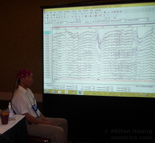
This picture is of a EEG demonstration with me wearing a red 19 channel EEG cap and the eyes open display on the screen.
EEG recordings usually require the subject to engage in different activities to be able to compare the different kinds of electrical activity the brain uses for those tasks. The most basic are sitting still with eyes closed or eyes open. Reading or math tasks are possible. In neurological seizure exams, strobe lights or hyperventilation might also be used. Perhaps the most specific type of test with the EEG is the examination for the ERP or event related potential. In this case, something might be flashed on a screen or a button click response might be required from the subject so that an exact timing of sensation or action can be compared to the EEG record to examine the related brain electrical response.
The complex data that is generated by the EEG can be analyzed using statistical procedures to look for patterns in particular frequencies or functional connections between different areas. The quantitative EEG or QEEG is a report of such statistics, and is sometimes referred to as a "brain map" for the visual presentation it uses.
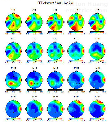
This is a 'brain map' of some of the activities at different frequencies, averaged over parts of the displayed EEG session. Such information can be compared to a database of age matched subjects to see if the electrical activity is more or less than 'normal.' This is one of the most common types of analysis performed in a QEEG. If such 'abnormal' activity correlates with clinical symptoms, it is possible to train the brain to reduce or increase its activity in the target location with the technique of neurofeedback.
- learn more about brainwaves - how they are generated and what they mean
- learn more about the different EEG machines
- learn more about types of EEG analysis and the QEEG
- learn more about ERPs (Event Related Potentials)